Magnetic resonance imaging
Magnetic resonance imaging, also known as magnetic resonance imaging (MRI), has become an indispensable part of modern medicine. Unlike computer tomography, the body is not exposed to radiation. This is because the sectional images are generated with the aid of a strong magnetic field, additional alternating fields, measuring antennas and a computer.
Magnetic resonance tomographic examinations ("MRI images") cannot replace X-ray and CT images in every respect, as they do not show bone structures in such detail and do not take biomechanics into account when the patient is lying down. However, they are often a very useful and necessary supplement to X-ray diagnostics, which is often performed in the standing position, in order to additionally reflect the real loading conditions.
Since an MRI examination usually produces a large number of cross-sectional images in different sequences, the diagnoses result from the sum of many individual images (quite possibly 100 or more).
In this respect, an MRI CD may contain more information than you are aware of.
According to the motto "4 eyes see more than 2", you can bring your MRI images and findings and we will analyze the content together.
I have been intensively involved with magnetic resonance imaging since its advent in the 1990s as part of my training at the Orthopedic University Hospital in Erlangen.
Although the clinic did not have its own MRI equipment at that time, we were already able to accompany patients through an arrangement with the Siemens company and examine them directly with Siemens latest equipment.
Even though we do not have our own MRI machine in our practice, we can arrange for a magnetic resonance imaging examination in your area at short notice.
Disc herniation with S1 root compression: Left before, right 8 weeks after 8 SpineMED® distractions

Complete Achilles tendon rupture

Disc prolapse L4/5 (lateral): left before, right after 15 SpineMED® distractions

Intervertebral disc sequestrum L1: Top before, bottom after 15 SpineMED® distractions

MRI-suitable pacemakers are now available, but here, too, the examination may only be performed under strict conditions and monitoring of the patient.
Metallic or magnetic objects such as EC or credit cards, cell phones, etc. have no place in the examination because the magnetic field damages them and pulls them into the device.
In addition, metallic objects can heat up and, in the worst case, cause burns.
Therefore, all objects containing metal must be removed before entering the examination room:
Patients who carry metal parts in their bodies must be sure to notify the medical staff in advance:
Newer metal implants are often made of non-magnetizable material such as titanium and are then unproblematic.Before the examination, however, it is imperative to check whether the implant is really suitable for MRI.
Magnetic resonance tomographic examinations ("MRI images") cannot replace X-ray and CT images in every respect, as they do not show bone structures in such detail and do not take biomechanics into account when the patient is lying down. However, they are often a very useful and necessary supplement to X-ray diagnostics, which is often performed in the standing position, in order to additionally reflect the real loading conditions.
Since an MRI examination usually produces a large number of cross-sectional images in different sequences, the diagnoses result from the sum of many individual images (quite possibly 100 or more).
In this respect, an MRI CD may contain more information than you are aware of.
According to the motto "4 eyes see more than 2", you can bring your MRI images and findings and we will analyze the content together.
I have been intensively involved with magnetic resonance imaging since its advent in the 1990s as part of my training at the Orthopedic University Hospital in Erlangen.
Although the clinic did not have its own MRI equipment at that time, we were already able to accompany patients through an arrangement with the Siemens company and examine them directly with Siemens latest equipment.
Even though we do not have our own MRI machine in our practice, we can arrange for a magnetic resonance imaging examination in your area at short notice.
Case studies
Disc herniation with S1 root compression: Left before, right 8 weeks after 8 SpineMED® distractionsComplete Achilles tendon rupture
Disc prolapse L4/5 (lateral): left before, right after 15 SpineMED® distractions
Intervertebral disc sequestrum L1: Top before, bottom after 15 SpineMED® distractions
Contraindications
MRI is usually not possible for patients with pacemakers or implantable cardiac defibrillators (ICDs), as damage can occur to the implant and also to the patient. The same applies to built-in insulin pumps or a cochlear implant - that is, an artificial inner ear.MRI-suitable pacemakers are now available, but here, too, the examination may only be performed under strict conditions and monitoring of the patient.
Metallic or magnetic objects such as EC or credit cards, cell phones, etc. have no place in the examination because the magnetic field damages them and pulls them into the device.
In addition, metallic objects can heat up and, in the worst case, cause burns.
Therefore, all objects containing metal must be removed before entering the examination room:
- Jewelry: (ear) rings, necklaces, bracelets, watches, piercings, hair clips
- Hearing aid, glasses, braces, removable dentures with metal parts
- Belts, buckles, clothing with buttons, metal zippers, underwire bras
- keys, pens, purses, coins
Patients who carry metal parts in their bodies must be sure to notify the medical staff in advance:
- Plates, screws, wires and staples after surgery
- artificial joint replacements/prostheses
- vascular stents
- fixed dentures
- IUD (contraception)
- artificial heart valves
- metal splinters (war, hunting, accident or explosion consequences)
- tattoos/permanent make-up with metallic colors
Newer metal implants are often made of non-magnetizable material such as titanium and are then unproblematic.Before the examination, however, it is imperative to check whether the implant is really suitable for MRI.
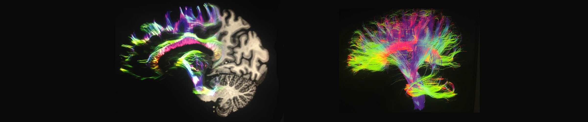
Diffusion MRI of the brain, half (Siemens company)

Specialist in Orthopaedics
Chiropractic
Sportsmedicine
Shockwave therapy
Acupuncture
Osteology
Naturopathic therapies